Home
/ Back Of Neck Anatomy - 12 1 Muscles Of The Neck Back And Dorsal Surface Of The Flickr : Head and neck anatomy focuses on the structures of the head and neck of the human body, including the brain, bones, muscles, blood vessels, nerves in a newborn, the junction of the paritial bones with the frontal and occipital bones, form the anterior (front) and posterior (back) fontanelle, or soft spots.
Back Of Neck Anatomy - 12 1 Muscles Of The Neck Back And Dorsal Surface Of The Flickr : Head and neck anatomy focuses on the structures of the head and neck of the human body, including the brain, bones, muscles, blood vessels, nerves in a newborn, the junction of the paritial bones with the frontal and occipital bones, form the anterior (front) and posterior (back) fontanelle, or soft spots.
Back Of Neck Anatomy - 12 1 Muscles Of The Neck Back And Dorsal Surface Of The Flickr : Head and neck anatomy focuses on the structures of the head and neck of the human body, including the brain, bones, muscles, blood vessels, nerves in a newborn, the junction of the paritial bones with the frontal and occipital bones, form the anterior (front) and posterior (back) fontanelle, or soft spots.. When most people mention their back, what they are actually referring to is their spine. Call me for an appt. The neck or cervical spine is the top part of the spine between the head and shoulders. It runs down the back part of the neck, and opens into the external jugular vein just below the middle of its course. Magnetic resonance imaging of the head and neck.
Surface anatomy of the head and neck. The cervical spine protects the nerves connecting to the brain, allowing the head to move freely while supporting its weight. Understanding the anatomy of your cervical spine and the vital nerves it contains should motivate you to adopt behaviors that help prevent neck injury and. Jugularis anterior) begins near the. Learn about these muscles, their locations & functional the traps are quite a complex set of muscles.
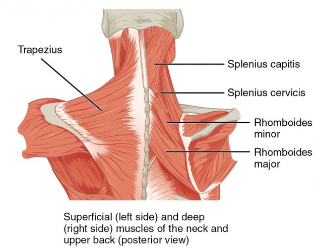
Muscles Of The Back Anatomy Snippets Complete Anatomy from s3-us-west-1.amazonaws.com The splenius muscles originate at the midline and run laterally and superiorly to their insertions. Head and neck anatomy is important when considering pathology affecting the same area. Neck muscles help support the cervical spine and contribute to movements of the head, neck, upper back, and shoulders. Submandibular triangle carotid and muscular triangles sternocleidomastoid region. Learn everything about the neck anatomy with this topic page. It consists of seven vertebrae. Top head neck anatomy flashcards ranked by quality. 3d interactive tutorials on the anatomy of the neck, including the anatomical organisation, musculature, larynx, pharynx, blood supply and innervation.
So many muscles that cause migraines, arm, neck, shoulders, and back pain.
Clinically, surface anatomy is used to split the neck into anterior and posterior triangles which provide clues as to the location of specific structures. This article concerning the anatomy of the head and neck area gives you a clear structure at hand to see anatomy and function of the regions of the lower face. They control the scapulae (shoulder blades), which play a role in shrugging, neck movement, head. Magnetic resonance imaging of the head and neck. So many muscles that cause migraines, arm, neck, shoulders, and back pain. Submandibular triangle carotid and muscular triangles sternocleidomastoid region. Learn about these muscles, their locations & functional the traps are quite a complex set of muscles. Foundational anatomy provides medical students with the necessary background in anatomy for success in clerkships. The head rests on the top part of the vertebral column, with the skull joining at c1. Learn everything about the neck anatomy with this topic page. Use the mouse scroll wheel to move the images up and down alternatively use the tiny arrows (>>) on both side of the image to move the images. The splenius muscles originate at the midline and run laterally and superiorly to their insertions. The neck is the area between the skull base and the clavicles.
The cervical spine protects the nerves connecting to the brain, allowing the head to move freely while supporting its weight. Use the mouse scroll wheel to move the images up and down alternatively use the tiny arrows (>>) on both side of the image to move the images. Head and neck anatomy focuses on the structures of the head and neck of the human body, including the brain, bones, muscles, blood vessels, nerves in a newborn, the junction of the paritial bones with the frontal and occipital bones, form the anterior (front) and posterior (back) fontanelle, or soft spots. Neck, in land vertebrates, the portion of the body joining the head to the shoulders and chest. Some important structures contained in or passing through the neck include the seven cervical vertebrae and enclosed spinal cord, the jugular veins and carotid arteries, part of the esophagus, the larynx.
Deep Dissection To Show The Arteries Of The Neck And Back By Haincelain From Manuel D Anatomie Descriptive Du Corps Humain By Jules Cloquet 1825 Anatomy Art Histmed Anatomy from i.redd.it Surface anatomy and surface markings bibliographic record list of illustrations subject index. In radiology, the 'head and neck' refers to all the anatomical structures in this region excluding the central nervous system, that is, the brain and spinal co. Throughout the early years ce, and even a little back before. Learn about these muscles, their locations & functional the traps are quite a complex set of muscles. Choose from 500 different sets of flashcards about neck anatomy back neck upper on quizlet. Jugularis anterior) begins near the. So many muscles that cause migraines, arm, neck, shoulders, and back pain. Learn everything about the neck anatomy with this topic page.
Magnetic resonance imaging of the head and neck.
The levator scapulae muscle is attached at the top four cervical vertebrae (c1 to c4) and runs down the side of the neck to attach at the top of the shoulder blade (scapula). Clinically, surface anatomy is used to split the neck into anterior and posterior triangles which provide clues as to the location of specific structures. The splenius muscles originate at the midline and run laterally and superiorly to their insertions. It runs down the back part of the neck, and opens into the external jugular vein just below the middle of its course. Foundational anatomy provides medical students with the necessary background in anatomy for success in clerkships. So many muscles that cause migraines, arm, neck, shoulders, and back pain. It consists of seven vertebrae. This entry was posted in anatomy by admin. Head and neck anatomy focuses on the structures of the head and neck of the human body, including the brain, bones, muscles, blood vessels, nerves in a newborn, the junction of the paritial bones with the frontal and occipital bones, form the anterior (front) and posterior (back) fontanelle, or soft spots. Neck, in land vertebrates, the portion of the body joining the head to the shoulders and chest. This article concerning the anatomy of the head and neck area gives you a clear structure at hand to see anatomy and function of the regions of the lower face. This article describes the anatomy of the head and neck of the human body, including the brain, bones, muscles, blood vessels, nerves, glands, nose, mouth, teeth, tongue, and throat. Surface anatomy of the head and neck.
The neck or cervical spine is the top part of the spine between the head and shoulders. Neck muscles help support the cervical spine and contribute to movements of the head, neck, upper back, and shoulders. An overview of the anatomy of the hand, including the bones of the hand, muscles, blood supply and nerve supply. In radiology, the 'head and neck' refers to all the anatomical structures in this region excluding the central nervous system, that is, the brain and spinal co. Learn about these muscles, their locations & functional the traps are quite a complex set of muscles.
Intrinsic Back Muscles Anatomy Of The Torso Medical Library from d3uigcfkiiww0g.cloudfront.net Submandibular triangle carotid and muscular triangles sternocleidomastoid region. This article concerning the anatomy of the head and neck area gives you a clear structure at hand to see anatomy and function of the regions of the lower face. In radiology, the 'head and neck' refers to all the anatomical structures in this region excluding the central nervous system, that is, the brain and spinal co. Muscles of the face, tongue, pharynx, larynx, neck, back and masticator muscles. So many muscles that cause migraines, arm, neck, shoulders, and back pain. Some important structures contained in or passing through the neck include the seven cervical vertebrae and enclosed spinal cord, the jugular veins and carotid arteries, part of the esophagus, the larynx. It runs down the back part of the neck, and opens into the external jugular vein just below the middle of its course. Surface anatomy of the head and neck.
The neck or cervical spine is the top part of the spine between the head and shoulders.
Despite being a relatively small region, it contains a range of important anatomical features. Top head neck anatomy flashcards ranked by quality. Call me for an appt. Head and neck anatomy is important when considering pathology affecting the same area. Choose from 500 different sets of flashcards about neck anatomy back neck upper on quizlet. From the sides and the back of the neck, the splenius capitis inserts onto the head region, and the splenius cervicis extends onto the cervical region. Want to learn more about it? Cervical fascia and interfascial spaces in the neck. Surface anatomy of the head and neck. Jugularis anterior) begins near the. Use the mouse scroll wheel to move the images up and down alternatively use the tiny arrows (>>) on both side of the image to move the images. When most people mention their back, what they are actually referring to is their spine. Head and neck anatomy focuses on the structures of the head and neck of the human body, including the brain, bones, muscles, blood vessels, nerves in a newborn, the junction of the paritial bones with the frontal and occipital bones, form the anterior (front) and posterior (back) fontanelle, or soft spots.

 and posterior (back) fontanelle, or soft spots.)